
Fig. S1
- ID
- ZDB-FIG-170815-10
- Publication
- Garcia et al., 2017 - Sheath Cell Invasion and Trans-differentiation Repair Mechanical Damage Caused by Loss of Caveolae in the Zebrafish Notochord
- Other Figures
- All Figure Page
- Back to All Figure Page
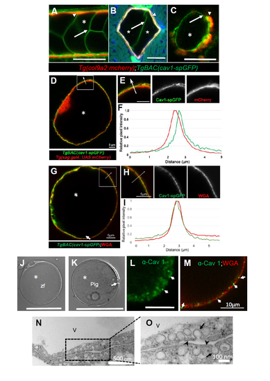
Caveolin 1 is a conserved vacuolated cell membrane protein in the vertebrate notochord. Related to Figure 1. A-C: Confocal images of a 72 hpf larva expressing col9a2:mcherry, BAC(cav1-spGFP). A: Live image. Arrows point to vacuolated cell membranes, asterisks mark vacuole lumen. B: Cross section. Arrows point to vacuolated cell membranes, asterisks mark vacuole lumen. C: Live image of a partially dissociated notochord showing that Cav1-GFP is expressed in both vacuolated and sheath cells. Arrowhead points to a sheath cell. D,F: Live image of a vacuolated cell isolated from a 48 hpf zebrafish embryo expressing BAC(cav1- spGFP) and sag:Gal4; UAS:mcherry-NTR;. Cav1-spGFP exhibits a punctate pattern that is separated from the cytoplasmic mcherry signal as shown by a line scan (F) along the arrow. G-I: Live image of a vacuolated cell isolated from a 48 hpf zebrafish embryo expressing BAC(cav1- spGFP) and stained with Alexa-594 WGA to label cell surface glycans. Cav1-spGFP exhibits a punctate pattern at the plasma membrane similar to that of WGA as shown by a line scan (I) along the arrow. J: DIC image of a vacuolated cell isolated from a 72 hpf zebrafish larva. Scale bar 50µm. K: DIC image of a vacuolated cell isolated from the nucleus pulposus (NP) of a 6 months old pig. Scale bar 50µm. L: Thick optical section confocal image of a portion of a pig vacuolated cell stained for Cav1. The antibody labels punctae at the cell surface. Arrows point to plasma membrane Cav1+ punctae. Scale bar: 5µm. M: Thin optical section confocal image of a portion of a pig vacuolated cell stained for Cav1 and WGA. N-O: Transmission electron micrographs of NP issue. Abundant caveolae (arrows) are present at the plasma membrane (arrow heads) in two neighboring cells. V marks a vacuole. |