
Fig. 4
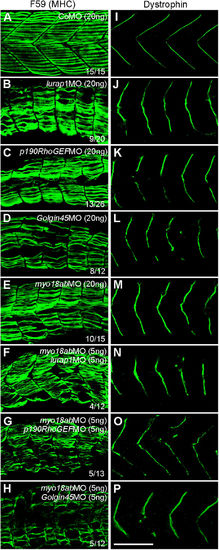
Functional interaction between MYO18A, Lurap1, p190RhoGEF and Golgin45 in myofiber integrity and dystrophin localization. Representative images showing the immunostaining results of slow muscle-specific MHC (A–H) and dystrophin (I–P) in control and various morphant embryos, as indicated. (A,I) Regular organization of myofibers within the somite (A) and localization of dystrophin at the sarcolemma (I) in CoMO-injected embryos, with clear chevron-shape myosepta. (B–E, J–M) Individual knockdown using high amounts of MOs against lurap1 (B,J), p190RhoGEF (C,K), Golgin45 (D,L) or myo18ab (E,M) produces similar muscle lesions (B–E) and disrupts dystrophin localization (J–M) at the sarcolemma. (F–H, N–P) Simultaneous knockdown using low amounts of indicated MOs also affects myofiber integrity (F–H) and dystrophin localization (N–P), with severely disorganized myofibers and strongly disrupted myosepta in a high proportion of morphant embryos. Scale bar: 100 μm. |
| Antibodies: | |
|---|---|
| Fish: | |
| Knockdown Reagents: | |
| Anatomical Terms: | |
| Stage: | Prim-5 |
| Fish: | |
|---|---|
| Knockdown Reagents: | |
| Observed In: | |
| Stage: | Prim-5 |