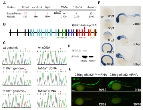
The fn10a locus encodes Eftud2 in zebrafish. (A) Regional fine-map of fn10a with the flanking genetic markers in chromosome 3, showing that eftud2 is located between genetic markers 10j-9 and 231?276. (B) A C-to-T mutation in exon 24 led to premature termination of eftud2 translation that produced mutant Eftud2 protein lacking 165 amino-acids in the C-terminus. (C) Sequencing data of genomic and cDNA of wild-type sibling, heterozygous, and mutant fn10a embryos. Note that mutant eftud2 mRNA is not stable in fn10a+/? mutants. (D) Western blot showing that Eftud2 protein was not detectable in fn10a?/? mutants. ?-Actin was used to normalize protein loading. (E) Left panels: TUNEL staining for neuronal apoptosis in only 12/68 fn10a mutants was rescued (lower panel) while that in 50/62 mutants was not rescued (upper panel) by mutant eftud2fn10a mRNA. Right panels: TUNEL staining for neuronal apoptosis in 39/48 fn10a mutants was rescued (lower panel) but that in 9/48 mutants was rescued less (upper panel) by wild-type eftud2 mRNA. All fn10a mutants were confirmed by PCR-based genotyping. (F) Whole-mount in situ hybridization revealed that eftud2 was broadly expressed at 3, 6, 12, 16 and 24 hpf and enriched in the brain and branchial arches at 36 and 48 hpf. Scale bar, 200 ?m.
|
