
Fig. S2
- ID
- ZDB-FIG-170516-29
- Publication
- Münch et al., 2017 - Notch signalling restricts inflammation and serpine1 expression in the dynamic endocardium of the regenerating zebrafish heart.
- Other Figures
- All Figure Page
- Back to All Figure Page
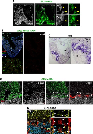
The injury endocardium exhibits filopodia-like protrusions and high levels of cdh5 (A) Vibratome section of ET33-mi60A heart, immune-stained for GFP and with phalloidin (7 dpci). Filopodia-like protrusions of wound endocardial cells show phalloidin staining (A', A'', arrowheads) (B) cdh5 FISH combined with IF showing low levels of cdh5 levels (red) in GFP+ endocardial cells all over the ventricle. (C) ISH against cdh5 at 7 dpci, showing strong expression within the injury site (is, arrow) and weak expression in the remote region (arrowheads). Boxed areas are magnified below or on the right. (D, E) IHC against GFP and Col1 on sections of ET33-mi60a transgenic hearts, showing low Col1 signal at 3 dpci (B) and similar distribution of endocardial cells and col1 at the inner injury border (dotted line) at 7dpci (C, white arrowheads). Scale bars: 50 µm in A, E; 200 µm in B, C; 100 µm in D, 25 µm in magnified views in A, E, 50 µm in B, C. |
| Gene: | |
|---|---|
| Fish: | |
| Condition: | |
| Anatomical Term: | |
| Stage: | Adult |