
Fig. 3
- ID
- ZDB-FIG-170125-12
- Publication
- Beretta et al., 2017 - Early Commissural Diencephalic Neurons Control Habenular Axon Extension and Targeting
- Other Figures
- All Figure Page
- Back to All Figure Page
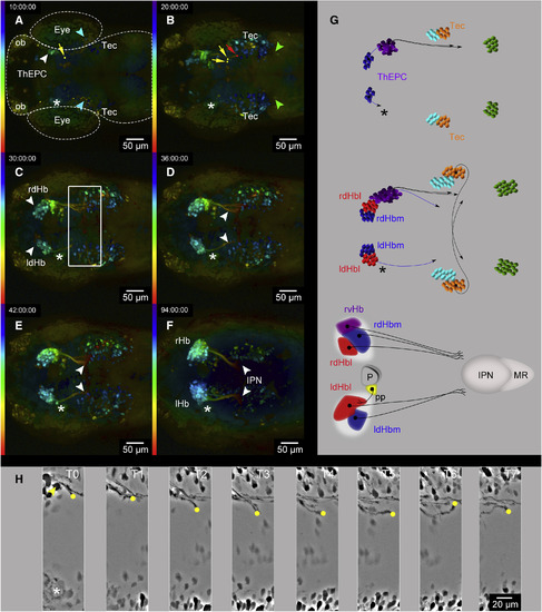
Unilateral ThEPC Cell Ablation Causes the Arrest of dHb Efferent Axons on Both Brain Hemispheres (A–F) Dorsal view, anterior to the left, color-coded MIP of six developmental stages acquired by in vivo 2-PM after complete unilateral ThEPC ablation at 32 hpf in an Et(−1.0otpa:mmGFP)hd1 transgenic embryo. Left: the LUT shows the z color-coded table according to the depth of each slice. Original stacks were cropped and gamma was adjusted to a value of 0.60 for display purposes. The head morphology has been roughly outlined in (A) for orientation. (A) The yellow dot and arrow mark the tip of a ThEPC axon. The blue arrowheads highlight the position of the second cluster of commissural neurons. (B) The yellow dots and arrows mark the tips of axons starting to migrate toward the midline, but which turn subsequently. The red dot and arrowhead label the tip of an ipsilateral ThEPC axon. The green arrowheads highlight the position of the third cluster of commissural neurons. (C) White arrowheads show the bilateral expression of GFP in the dHb. A white square highlights the area in which dHb axon elongation stalls. Note the delay of dHb axons on the left side. (D and E) White arrowheads highlight the position of extending dHb efferent axons at (D) 68 hpf and (E) 74 hpf. (F) Architecture of the habenular neural circuit after left-sided ThEPC cell ablation at 5 dpf. White arrowheads mark the ends of the stalled dHb efferent axon bundles. (G) Summary of events during habenular neural circuit development after complete unilateral ThEPC ablation between (top to bottom) 42 and 52 hpf, 42 and 68 hpf, and 42 hpf and 5 dpf. ThEPCs and rvHb, purple; Tec, orange; second cluster of projection neurons, light blue; third cluster of projection neurons, green; ldHb/rdHb and axonal projections, blue/red. (H) Dorsal view, anterior to the left, MIPs of eight developmental stages focused on a turning commissural ThEPC axon on the non-ablated site. The axon’s tip is marked by a yellow dot and its neuron with a yellow arrowhead. Segmented images show events between 34 and 43 hpf. MIPs were adjusted using the difference of Gaussians to better visualize the structure of interest. Asterisks mark the site of ThEPC ablation between 32 hpf and 5 dpf. d, dorsal; Hbl, lateral habenula; Hbm, medial habenula; IPN, interpeduncular nucleus; l, left; MR, median raphe; ob, olfactory bulb; r, right; Tec, optic tectum; ThEPC, thalamic-epithalamic early projecting cluster; v, ventral. See also Figures S3 and S4 and Movie S3. |