
Fig. 3
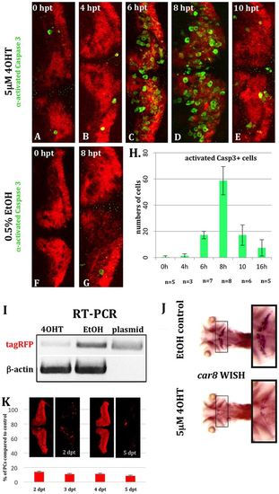
Time course of apoptosis in PC-ATTACTM larvae. (A-G) Whole-mounts of 5-dpf transgenic PC-ATTACTM embryos were stained with an antibody against activated Caspase 3 (green) after treatment at 4 dpf with 5×10⁻⁶M 4OHT (A=0 h; B=4 h; C=6 h; D=8 h; E=10 h) or 0.5% EtOH (F=0 h; G=8 h). (H) Significant elevation of apoptosis could be observed after 6 h, peaking after 8 h of 4OHT treatment based on PC counts at 4 dpf. (I-K) To score the extent of PC ablation over time, we performed 4OHT treatments with 4 dpf heterozygous larvae for 16 h (compared with 0.5% EtOH controls) and analyzed the cell death 24 h later. (I) RT-PCR revealed a strong reduction of fyntagRFP mRNA for 4OHT-treated larvae in contrast to EtOH controls with β-actin used as ubiquitously expressed control gene. (J) Although mRNA in situ hybridization (n=5) showed carbonic anhydrase 8 (ca8) expression was prominent in the cerebellum in control larvae, virtually no ca8 expression was visible in PC-ablated specimens (black squares). (K) PC counts 2 and 5 days post-treatment (dpt) revealed a 90% reduction in PC number compared with EtOH controls (K). hpt, hours post-treatment. |