
Fig. 8
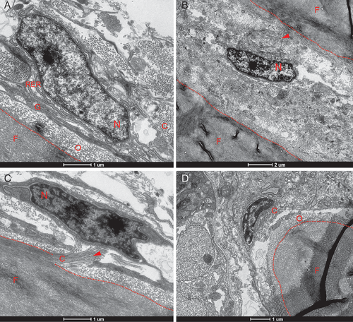
Transmission electron micrographs show various osteogenic cells within the interfrontal sutures. The calvarium was isolated from adult zebrafish at 14 wpf. (A) An osteoblast in direct contact with the osteoid of the upper frontal plate, indicative of ongoing ossification. (B) A mid-suture osteoblast not attached to bone. (C) The longitudinal organization of collagen fibrils along the frontal bone. (D) The tip of the lower plate of the frontal bone, with adjacent associated osteoblast. Abbreviations: C—collagen fibrils, F—frontal bone, G—Golgi apparatus, N—nucleus, O–osteoid, RER–rough endoplasmic reticulum; arrows indicate collagen fibrils. |