
Fig. S5
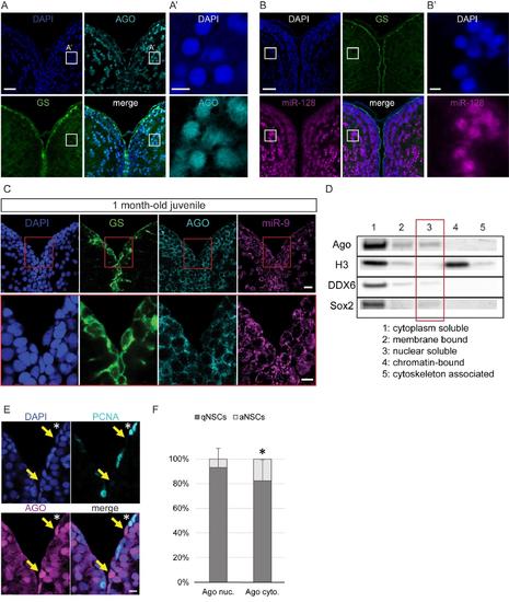
Nuclear localization of Argonaute proteins in the adult zebrafish pallium and its association with quiescence in mature NSCs, related to figure 4 (A-A’) Fluorescent immunodetection of Ago (light blue), GS (green), with DAPI counterstaining (dark blue). A’ is a zoom on the region indicated by a white square in A, showing a strong nuclear staining for AGO in some parenchymal neurons (B-B’) ISH using a LNA probe for miR-128 (magenta) combined with immunostaining for GS (green) and DAPI counterstaining (dark blue). B’ is a zoom on the region indicated by a white square in B, showing a strong nuclear staining for miR-128 in some parenchymal neurons. Scale bars=20μm (A,B) and 5μm (A’,B’) (C) Sections through the medial region of the dorsal telencephalon of a 1mpf juvenile fish showing ISH of miR-9 (magenta) and double immunofluorescence of GS (green) and Ago (light blue) with nuclear DAPI counterstaining (dark blue) demonstrating that miR-9 and Ago are located exclusively in the cytoplasm of the RG progenitors. Scale bars=10μm. (D) Western blot analysis of fractionated proteins samples from the adult zebrafish telencephalon. Substantial levels of Ago proteins can be detected in the nuclear soluble fraction (lane 3). (E) Double immunofluorescence of PCNA (light blue) and Ago (magenta) with a nuclear DAPI counterstaining (dark blue) illustrating that strong nuclear Ago is never in PCNA+ aNSCs (yellow arrows) and aNSCs express Ago in the cytoplasm (white asterisk). Scale bar=10μm. (F) Proportion of qNSCs (gfap+, MCM5-; dark grey) and aNSCs (gfap+, MCM5-; light grey) among NSCs with mostly nuclear (Ago nuc.) or cytoplasmic Ago (Ago cyto.) demonstrating an association between nuclear Ago and quiescence (χ2 p-value<0.01). n=3 brains; data are represented as mean ± 95% CI. |