
Fig. 3
- ID
- ZDB-FIG-161017-3
- Publication
- Hosseini et al., 2016 - Efferocytosis and extrusion of leukocytes determine the progression of early mycobacterial pathogenesis
- Other Figures
- All Figure Page
- Back to All Figure Page
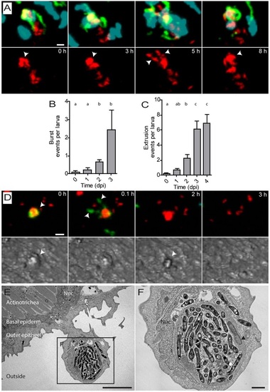
Macrophages containing aggregates of Mm undergo burst or extrusion. (A) Representative images from live-cell imaging of a large aggregate of Mm (arrowhead) inside a macrophage undergoing cell death at t=3h. Spreading of Mm was observed at t=5h. The aggregate was compacted again by newly recruited leukocytes at t=8h (see Movie 6). (B) Number of burst events observed at different time points after Mm infection in the tail fin. (C) Number of extrusion events observed at different time points after Mm infection in the tail fin. (D) Representative images from live-cell imaging showing an extrusion event for accumulated Mm in a macrophage (arrowheads). First, the macrophage undergoes cell death and subsequently after 3h the bacterial content is extruded. (E) TEM image of an extruding epithelial cell from the tail fin surrounding a dead cell containing a large aggregate of Mm. The two epithelial layers and actinotrichia are indicated. (F) Higher magnification of the region indicated in E, showing compact aggregates of Mm (asterisks) in the dead cell with a condensed nucleus (arrowhead). Error bars indicate s.e.m. (n~20 larvae per time point), means with the same letter do not differ significantly (Dunnett′s post-test, P<0.05). Scale bars: 10µm (A,D,E); 2µm (F). |
| Genes: | |
|---|---|
| Fish: | |
| Condition: | |
| Anatomical Terms: | |
| Stage: | Protruding-mouth |