
Fig. 6
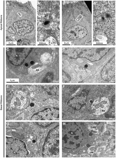
Ultrastructural features of ribbons during hair cell regeneration. (A-I) 5dpf larvae were treated with 400µM neomycin for 1h to kill all hair cells. To assess ribbon morphology during regeneration, larvae were fixed and processed for TEM and anterior lateral line neuromasts were imaged at different times post treatment (hpt). (A-B′) Electron-dense structures indicative of ribbon precursors are found in the cytoplasm of hair cells at early stages (18hpt and 24hpt) during regeneration. (A′,B′) Close up views of boxed regions in A,B. (C-H) Basal ribbons at different stages post treatment. Immature ribbons are seen at early stages such as double ribbons at 12hpt (C) and ‘ectopic’ or ‘floating’ ribbons at 24hpt (E) [the ribbon faces a support cell process (asterisk), which still separates the hair cell from a nearby afferent ending]. The immature ribbon anchored at the membrane at 48hpt (G) faces a growth cone (asterisk), which has the characteristic dense cytoplasm filled with different size vesicles. Mature synapses (D,F,H) were clearly seen from 24hpt to 48hpt. N, nucleus; EN, efferent nerve ending; AN, afferent nerve ending. The images in D-H are shown at the same scale as that in C. |