
Fig. 4
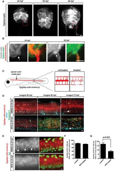
Radial glial cells are specifically ablated in Tg(gfap:nsfB-mcherry) embryos. A: Images from Tg(gfap:egfp) embryos at 24, 36, and 48 hpf. Box denotes location of MEP TZ shown in B. B:. Zoomed images of Tg(gfap:egfp) embryos stained with an antibody to acetylated tubulin to mark motor axons. Arrow denotes location of MEP. C: Schematic of Tg(gfap:nsfB-mcherry) embryo with zoomed-in views of the spinal cord in untreated and MTZ-treated animals showing death (dashed lines) of radial glial cells in MTZ-treated animals. D: Confocal images of Tg(gfap:nsfB-mcherry) animals treated with DMSO or MTZ at 24 hpf and fixed and stained with antibodies specific to GFAP and Caspase-3 at 36, 48, and 72 hpf. Arrow denotes floorplate radial glia that do not express NTR and are present after MTZ-treatment. E: Confocal images of Tg(gfap:nsfB-mcherry) animals treated with DMSO or MTZ at 24 hpf and fixed and stained with antibodies specific to Isl1 and Sox10 at 56 hpf. Images show mcherry+ endfeet (denoted with arrows) in control animals that are absent in MTZ-treated animals. Quantification of (F) Isl1+ motor neurons (G) and Sox10+ glia in the spinal cord at 56 hpf in DMSO and MTZ-treated embryos. Scale bars, 25 µm. |