
FIGURE
Fig. 9
- ID
- ZDB-FIG-160602-19
- Publication
- McCarthy et al., 2016 - Pdgfra and Pdgfrb genetically interact during craniofacial development
- Other Figures
- All Figure Page
- Back to All Figure Page
Fig. 9
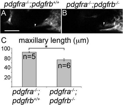
Maxillary domain CNCC length is reduced in pdgfra;pdgfrb mutants at 30 hpf. A,B: Thirty hpf single z-stack confocal images of fli1:EGFP transgenic embryos with indicated genotypes above the panels, anterior is to the left. EGFP-labeling is depicted as gray. The maxillary condensation extends further anteriorly in (A) pdgfra-/-;pdgfrb+/+ embryos compared with (B) pdgfra-/-;pdgfrb-/- embryos (arrowhead denotes length measured). C: Bar chart depicting maxillary neural crest length (Students t-test; *P = 0.0136). Scale bar = 50 µm. |
Expression Data
| Gene: | |
|---|---|
| Fish: | |
| Anatomical Term: | |
| Stage: | Prim-15 |
Expression Detail
Antibody Labeling
Phenotype Data
| Fish: | |
|---|---|
| Observed In: | |
| Stage: | Prim-15 |
Phenotype Detail
Acknowledgments
This image is the copyrighted work of the attributed author or publisher, and
ZFIN has permission only to display this image to its users.
Additional permissions should be obtained from the applicable author or publisher of the image.
Full text @ Dev. Dyn.