
Fig. 4
- ID
- ZDB-FIG-160205-50
- Publication
- Cronan et al., 2015 - CLARITY and PACT-based imaging of adult zebrafish and mouse for whole-animal analysis of infections
- Other Figures
- All Figure Page
- Back to All Figure Page
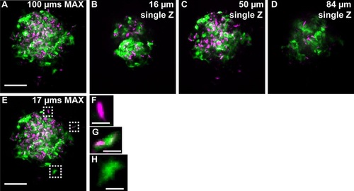
Fluorescent mycobacteria and cytokine induction can be imaged deep within intact adult zebrafish. (A) The TNF reporter (green) is expressed throughout a large granuloma (tdTomato-expressing M. marinum: magenta) in the TgBAC(tnf:GFP) line. Imaging begins 256µm below the scales and the stack (A) is µ100µm deep; individual Z planes from the stack (B-D) reveal TNF reporter intensity throughout the granuloma. (E-H) TNF reporter expression in the granuloma is not dependent on infection status of individual cells: (F) an infected cell that does not express the TNF reporter; (G) an infected cell expressing the TNF reporter; (H) an uninfected cell expressing the TNF reporter. Scale bars: 50µm (A-E); 5µm (F-H). Single Z frames were exported and gamma adjusted in FIJI/ImageJ for increased visibility, with all gamma adjustments applied uniformly across all images. |