
Fig. 1
- ID
- ZDB-FIG-160205-47
- Publication
- Cronan et al., 2015 - CLARITY and PACT-based imaging of adult zebrafish and mouse for whole-animal analysis of infections
- Other Figures
- All Figure Page
- Back to All Figure Page
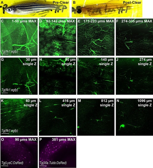
CLARITY protocol adapted for imaging intact zebrafish adults. (A,B) Zebrafish adult pre- and post-clearing. (C-N) Whole-body CLARITY allows imaging of fluorescent vasculature deep within the adult body. Blood vessels labeled by Tg(flk1:egfp) are imaged from the surface to deep within while maintaining fluorescence intensity and resolution. (C-J) Individual images obtained using an SP8 confocal microscope, ranging from the animal′s scales (surface=1µm) to 335µm deep. (C-F) Z-stack is split into ~50µm maximum projection images to allow for clear views of vascular structures. (G-J) Individual Z planes from stack. (K-N) Individual images from two-photon microscopy ranging from the animal′s scales (surface=1µm) to >1mm deep. 1-µm optical sections are shown. Scale bars: 100µm. Single Z frames were exported and gamma adjusted in FIJI/ImageJ for increased visibility, with all gamma adjustments applied uniformly across all images. (O,P) CLARITY techniques are compatible with red fluorescent proteins. (O) Neutrophils within the epidermis were imaged using the transgenic line Tg(LysC:DsRed). 90-µm maximum projection image. (P) Neuronal cell bodies within the eye of cleared zebrafish in a 381-µm maximum projection from the transgenic line Tg(Xla.Tubb:DsRed). |