|
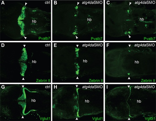
Immunostaining with (A-C) anti-Pvalb7 and (D-F) anti-zebrinII antibody show loss of cerebellar Purkinje cells in atg4da morphants. (A,D) Control embryos at 4.5 dpf. (B,E) Morphants with mild phenotype show partial loss of cerebellar Purkinje cells. (C,F) Morphants with strong phenotype show either total loss of Purkinje cells or presence of few differentiated neurons, which are laterally located in the cerebellum. (G-I) Labeling of cerebellar granule cells with anti-Vglut1 antibody in control and morphant embryos. (H) Mildly affected morphants show reduced expression of Vglut1 in the cerebellum. (I) Vglut1 expression is strongly reduced in embryos showing severe phenotype. White arrowheads indicate the region of cerebellum. Abbreviations: hb, hindbrain.
|