
Fig. 2
- ID
- ZDB-FIG-150406-27
- Publication
- Boer et al., 2015 - Fascin1-Dependent Filopodia are Required for Directional Migration of a Subset of Neural Crest Cells
- Other Figures
- All Figure Page
- Back to All Figure Page
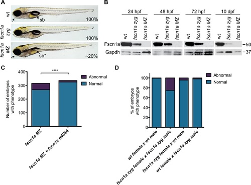
Maternal Fscn1a protein persists throughout embryogenesis and masks morphological defects in fscn1a zygotic mutants. (A) Brightfield images of 5 dpf wild type (wt), zygotic fscn1a (fscn1a zyg), and maternal/zygotic fscn1a (fscn1a MZ) embryos. Arrow highlights craniofacial cartilage, sb = swim bladder. Number indicates percentage of embryos with phenotype. (B) Immunoblot showing Fscn1a protein levels at 24, 48, 72 hpf and 10 dpf in wt, fscn1a zyg, and fscn1a MZ mutant embryos. Numbers on right denote molecular mass markers. (C) Quantitation of uninjected or fscn1a mRNA-injected fscn1a MZ embryos with normal vs. abnormal craniofacial cartilages at 5 dpf (n = 315 fscn1a MZ embryos, 336 fscn1a MZ + fscn1a mRNA embryos, ****p<0.0001). (D) Quantitation of embryos with normal vs. abnormal craniofacial cartilages at 5 dpf (n = 201 wt female × wt male, 119 fscn1a zyg female × fscn1a zyg male, 115 fscn1a zyg female × wt male, 151 wt female × fscn1a zyg male). |
| Fish: | |
|---|---|
| Observed In: | |
| Stage: | Day 5 |