
Fig. 4
- ID
- ZDB-FIG-150211-21
- Publication
- Paulus et al., 2014 - Loss of Optineurin In Vivo Results in Elevated Cell Death and Alters Axonal Trafficking Dynamics
- Other Figures
- All Figure Page
- Back to All Figure Page
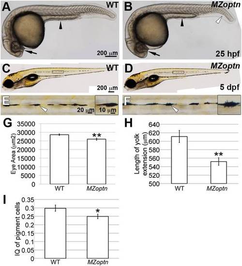
MZoptn embryos have subtle morphological defects at early developmental ages. A-F. Representative images of live WT (A, C, E) or MZoptn (B, D, F) embryos at 25 hpf (A, B) or 5 dpf (C-F). Anterior is to the left. A, B. The area of the eyes (arrows) and length of the yolk tube extension (arrowheads) are smaller in MZoptn embryos. MZoptn embryos occasionally have a curve to the tail (open arrowhead). C-F. Pigment cells in MZoptn embryos have more filopodia. E and F are higher magnification views of boxed areas in C and D. Insets in E and F are higher magnifications of pigment cells indicated by arrows. G-I. Quantification of eye size at 25 hpf (I), length of yolk tube extension at 25 hpf (J) and isoperimetric quotient of pigment cells at 5 dpf (K). * indicates p≤0.05, ** indicates p≤0.01. |
| Fish: | |
|---|---|
| Observed In: | |
| Stage Range: | Prim-5 to Day 5 |