
Fig. 5
- ID
- ZDB-FIG-141125-6
- Publication
- Choksi et al., 2014 - Systematic discovery of novel ciliary genes through functional genomics in the zebrafish
- Other Figures
- All Figure Page
- Back to All Figure Page
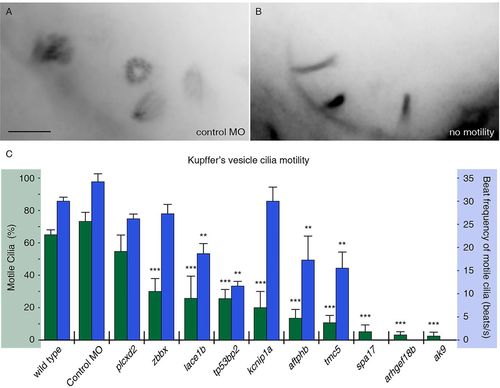
FIGs are required for ciliary motility. (A,B) Embryos were injected with RNA encoding Arl13b-GFP to label KV cilia and imaged using high framerate video microscopy. (A) An overlay of 20 frames from a movie from a control embryo indicates four moving cilia in view. (B) By comparison, overlays from movies of an embryo with immotile cilia show no blurring, indicating stationary objects. Scale bar: 5μm. (C) Measurements of ciliary motility in wild-type and morphant embryos targeting ten selected genes. Two aspects of ciliary motility were scored: the percentage of KV cilia that exhibited motility (green) and the average beat frequency of motile cilia (blue). Error bars represent s.e.m. ***P<4.0×10⁷ (Fisher′s Exact test, two-tailed; P-values are listed in supplementary material Table S3). **P<5.0×10³ (Student′s t-test, two-tailed, P-values are listed in supplementary material Table S3). The three genes without reported ciliary beat frequency did not have sufficient motile cilia for analysis (n<8). |
| Fish: | |
|---|---|
| Knockdown Reagents: | |
| Observed In: | |
| Stage: | 10-13 somites |