
Fig. 6
- ID
- ZDB-FIG-141024-6
- Publication
- Xu et al., 1995 - Expression of truncated Sek-1 receptor tyrosine kinase disrupts the segmental restriction of gene expression in the Xenopus and zebrafish hindbrain
- Other Figures
- All Figure Page
- Back to All Figure Page
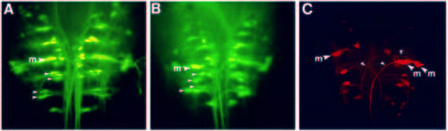
Effect of truncated Sek-1 on reticulospinal neurons in the zebrafish embryo. Reticulospinal neurons were revealed in 3 day zebrafish embryos by retrograde labelling from the spinal cord using LRD (A) Uninjected embryo. (B,C) Embryos injected with RNA encoding truncated Sek-1. The small arrowheads in A and B indicate the locations of corresponding pairs of reticulospinal neurons in r5 and r6. The spacing of these neurons is altered in the injected embryo shown in B. The small arrowheads in C indicate the axons of the duplicated Mauthner neurons in r4. The large arrows labelled ‘m’ indicate the Mauthner neuron. Scale bar, 50 µm. |
| Gene: | |
|---|---|
| Antibody: | |
| Fish: | |
| Anatomical Terms: | |
| Stage: | Prim-5 |