
Fig. 7
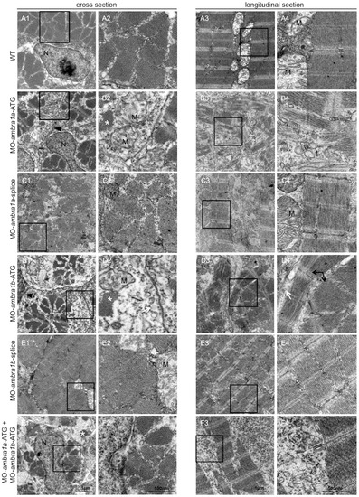
Ultrastructural analysis of ambra1 morphants muscles reveals disorganized sarcomeres. Representative electron micrographs of cross and longitudinal sections of 3 dpf (WT, panels A1–A4), ambra1a ATG-morphant (panels B1-B4), ambra1a splice-morphant (panels C1–C4), ambra1b ATG-morphant (panels D1–D4), ambra1b splice-morphant (panels E1-E4), and co-injected morphant (panels F1–F4) zebrafish embryos. Columns 2 and 4 show higher magnification views of the boxed areas in column 1 and 3, respectively. Muscles of WT and 5 m-control (not shown) embryos display well-organized myofibers, showing densely packed sarcomeres with regular organization of thin and thick myofilaments. ambra1 depleted muscles show a number of ultrastructural defects, with small patches of disorganized myofibers and mitochondria scattered throughout the cytoplasm. Black arrows, area with myofibrils having different orientations; white arrow, dilated sarcoplasmic reticulum not in contact with myofibrils; asterisks, fragments of torn myofibrils; M, mitochondria; N, nucleus. |
| Fish: | |
|---|---|
| Knockdown Reagents: | |
| Observed In: | |
| Stage: | Protruding-mouth |