
Fig. 6
- ID
- ZDB-FIG-140807-67
- Publication
- Danilova et al., 2014 - The role of DNA damage response in zebrafish and cellular models of Diamond Blackfan Anemia
- Other Figures
- All Figure Page
- Back to All Figure Page
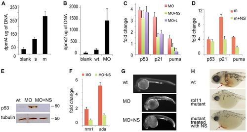
Rescue of RP-deficient zebrafish with exogenous nucleosides. (A) At 48 hpf, Rpl11 mutants incorporated more [3H] deoxycytidine into DNA than their wild-type siblings (P<0.002). The count adjusted to 4 μg of genomic DNA is shown. (B) At 48 hpf, embryos that had been injected with a morpholino against Rps19 incorporated more [3H]deoxycytidine into DNA than wild-type embryos (P<0.005). The count adjusted to 2 μg of genomic DNA is shown. (C) Supplementation with exogenous nucleosides (NS) decreased the expression of tp53, p21 and puma in zebrafish embryos that had been injected with a morpholino against Rps19 (MO). L, treatment with 0.5 mg/ml leucine was used for comparison. RT-qPCR analyses were performed at 22 hpf. The fold change in gene expression was calculated relative to expression in wild-type embryos. (D) The addition of exogenous nucleosides reduced the expression of tp53, p21 and puma in Rpl11-mutant zebrafish embryos. RT-qPCR analyses were performed at 48 hpf. The fold change in expression was calculated relative to expression in wild-type siblings. (E) Nucleoside treatment decreased the level of p53 protein in Rps19-deficient zebrafish embryos. 22 hpf. Western blot. (F) The addition of exogenous nucleosides normalized the altered expression of genes that are involved in nucleotide metabolism – rrm1 and ada. RT-qPCR analyses were performed at 22 hpf. In A–D and F, means±s.d. are shown. (G) Treatment with nucleosides decreased the amount of apoptosis. Embryos were injected with a morpholino against Rps19, and half of these were treated with nucleosides. Representative images are shown of fish at 28 hpf. Acridine orange staining shows fewer apoptotic cells in morphants that had been treated with nucleosides. (H) The addition of exogenous nucleosides increased the amount of red blood cells in Rpl11 mutants. Representative images are shown of O-dianizidine staining of fish at 80 hpf. Rpl11 mutants were confirmed by genotyping after staining. Arrows point to erythroid cells. |
| Fish: | |
|---|---|
| Knockdown Reagent: | |
| Observed In: | |
| Stage Range: | Prim-5 to Protruding-mouth |