
Fig. 2
- ID
- ZDB-FIG-140807-26
- Publication
- Koole et al., 2014 - Mosaic analysis and tumor induction in zebrafish by microsatellite instability-mediated stochastic gene expression
- Other Figures
- All Figure Page
- Back to All Figure Page
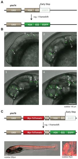
UAS-reporters stochastically express H2A-EGFP. (A) (Upper panel) Schematic representation of reporter plm74 in which frameshifts at a G23 microsatellite can result in EGFP expression. Note that, for illustration purposes, we used the example of a 1 frameshift, however other, bigger, frameshifts can also lead to in-frame EGFP. UAS, upstream activator sequences. (B) Time-lapse images of the head region of an embryo derived from crossing Et(E1b:Gal4-VP16)s1101t fish to Tg(UAS:H2A-G23-EGFP)hu6243 fish. Stochastic expression of nuclear EGFP (green) in cells was observed, and individual cells and their descendants can be traced over time (denoted by the letters A-F in the images). Images 1–4 represent pictures taken around 16, 21, 25 and 31 hpf and correspond to supplementary material Movie 1. (C) (Upper panel) Schematic representation of reporter plm76 in which Myr-TdTomato is placed downstream of a UAS-cassette, followed by a T2A sequence and H2A-G23-EGFP, meaning that EGFP is out-of-frame. Transgenic F1 animals [Et(E1b:Gal4-VP16)s1101t fish crossed to Tg(UAS:Myr-TdTomato-H2A-G23-EGFP)hu6242] exhibit cells that express fluorescent membrane-labeled TdTomato and, in a mosaic pattern caused by in vivo stochastic frameshifting, EGFP-fluorescent nuclei (lower panels, an enlarged image of the boxed area is shown in the right-hand image). |