
Fig. 6
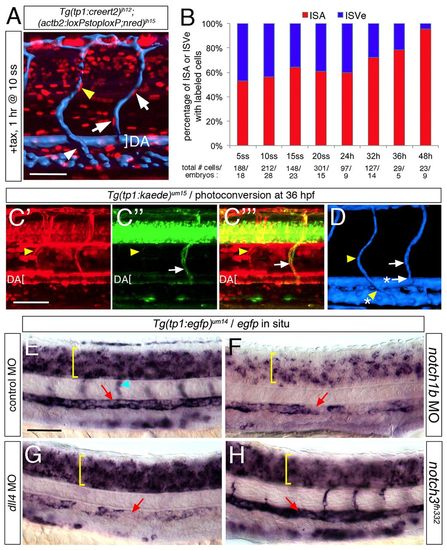
Notch is downregulated in ISVs during arteriovenous re-programming. (A) Two-photon imaging following microangiography (surface-rendered in blue) of Tg(tp1:creert2)jh12;(actb2:loxP-stop-loxP;nred)jh15 embryo at 60 hpf following exposure to tamoxifen at 10 ss for 1 hour. Yellow arrowhead denotes red endothelial nucleus lining an intersomitic vein, as determined by its connection to the posterior cardinal vein (white arrowhead). White arrows indicate cells lining an intersomitic artery connected to the dorsal aorta (indicated by white bracket). (B) Graph showing proportion of intersomitic arteries or veins with red fluorescent cells in Tg(tp1:creert2)jh12;(fli1ep:loxPnBlueloxP;red)um43 embryos at 60 hpf following a 1 hour tamoxifen pulse at the indicated stage; total numbers of labeled cells and embryos counted are indicated below the x-axis. (C,D) Two-photon micrographs of same Tg(tp1:kaede)um15 embryo at 60 hpf following photoconversion of Kaede at 36 hpf. Yellow arrowheads denote intersomitic vein endothelial cell positive for red, but not green fluorescence. White arrow denotes an intersomitic artery endothelial cell that maintained Kaede expression. (C) Red fluorescence indicating cells that exhibited Notch activation upon photoconversion at 36 hpf. (C2) Green fluorescence from Kaede expressed after photoconversion. (C2) Overlay of images in C and C2. (D) Surface rendering of image stack following microangiography of embryo in C; asterisked arrowhead and arrow denote connection to posterior cardinal vein and dorsal aorta, respectively. (E-H) Tg(tp1:egfp)um14 embryos at 30 hpf subjected to whole-mount in situ hybridization with an egfp riboprobe. Embryos injected with 2.5 ng of control MO (E), 2.5 ng of notch1b MO (F), 15 ng of dll4 MO (G) or mutant for notch3fh332 (H). Yellow bracket denotes neural tube, light blue arrowhead indicates the intersomitic vessels, and red arrow indicates the dorsal aorta. Scale bars: 50 μm. |
| Genes: | |
|---|---|
| Fish: | |
| Condition: | |
| Knockdown Reagents: | |
| Anatomical Terms: | |
| Stage Range: | Prim-15 to Pec-fin |