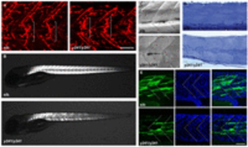
Muscle integrity is impaired in y241 mutants. (A) α-bungarotoxin-conjugated Alexa-Fluor-555 staining of 2 dpf wild-type sibling (left) and y241 mutant (right) embryos, showing equatorially located neuromuscular junctions (brackets). Scale bar: 50 μm. (B) Birefringence in live 5 dpf wild-type sibling (top) and y241 mutant (bottom) larvae detected using polarized light. Mutants show reduced and patchy birefringence. (C) Dodt-gradient-contrast illumination of muscle in 3 dpf larvae after removal of skin. Internal muscle striations are visible in myofibers stretching between somite boundaries in siblings (top) but not in y241 mutants (bottom). (D) 10 μm toluidine-blue-stained sections through the tail of 3 dpf wild-type siblings (top) and y241 mutants (bottom). (E) Kaede expression in highly variegated 24 hpf y241; UAS:kaede sibling embryos (top panels) and mutants (bottom panels) with DAPI staining to reveal nuclear positions. Somite boundaries observed in bright-field images are indicated by dashed lines. Scale bar: 30 μm.
|
