
Fig. 2
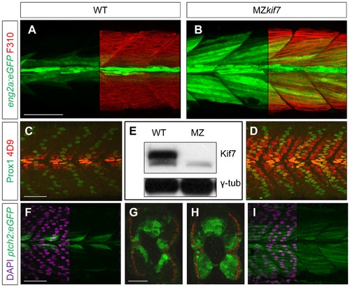
Absence of Kif7 protein and de-repression of Hh target genes in MZkif7 embryos. (A,B) Lateral images of 30 hpf wild-type (A) and MZkif7 mutant (B) embryos expressing eng2a:eGFP (green); note dramatic expansion of eng2a:eGFP expression within the fast-twitch fibers revealed by F310 staining (red) in the merged image (right panel). (C,D) Parasagittal optical sections of wild-type (C) and MZkif7 mutant (D) 30 hpf embryos stained with anti-Prox1 (green) and mAb4D9 (red) (E) Western blot of wild-type (WT) and MZkif7 mutant (MZ) embryo extracts probed with polyclonal rabbit anti-Kif7 antiserum showing complete loss of Kif7 protein (upper band) from MZkif7 embryos. Loading control: γ-tubulin (γ-tub). (F,I) Parasagittal and (G,H) transverse optical sections of 30 hpf wild-type (F,G) and MZkif7 (H,I) embryos expressing a ptch2:eGFP transgene (green) showing the expansion of the ptch2 expression domain in the myotome and neural tube in the absence of Kif7. Nuclei are revealed in left half (of panels F,I) by DAPI staining (purple). The edge of the myotome is shown by mAbF9 (in G,H) marking the superficial slow-twitch muscle fibers. Scale bar: 50 μm. |
| Genes: | |
|---|---|
| Antibodies: | |
| Fish: | |
| Anatomical Terms: | |
| Stage: | Prim-15 |
| Fish: | |
|---|---|
| Observed In: | |
| Stage: | Prim-15 |