|
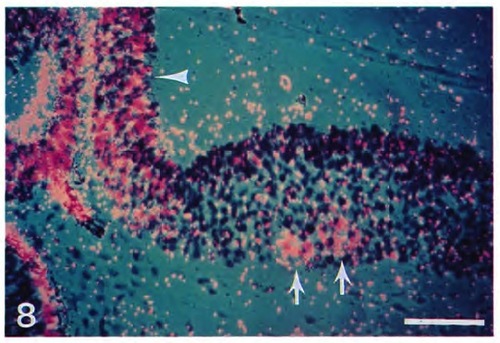
Cells in the first three hindbrain rhombomeres express the eng3 gene. A parasagittal section through the hindbrain of a 32 h embryo was hybridized with a probe for the eng3 gene. Small clusters of cells (arrows) in the ventral parts of the second and third rhomobomeres hybridize. A cluster of cells in the first rhombomere (contained in a different section, not shown, and as illustrated by the whole-mount embryo in Fig. 6 H) also hybridized. The hybridization at the junction between the midbrain and hindbrain is also apparent (arrowhead). The signal in the lower left is caused by the pigment epithelium of the retina and is not due to specific hybridization as shown in unhybridized control sections. Scale bar, 50 μm.
|