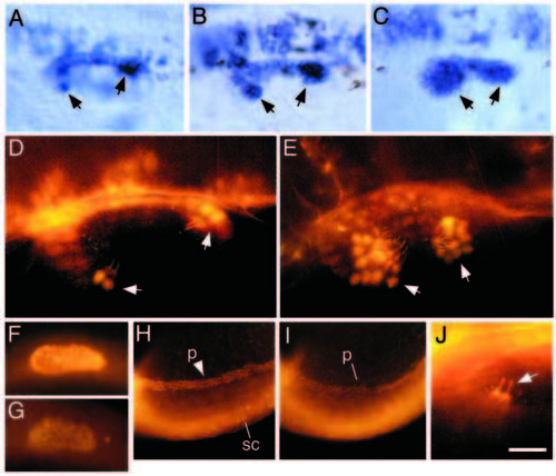
Expression of pax2.1 in the inner ear of wildtype and dlAdx2/dx2 embryos. (A-C) Dorsolateral view of the otic vesicle at 24 h showing accumulation of pax2.1 transcripts in a wild-type embryo (A), a moderately affected dlAdx2/dx2 mutant (B) and a severely affected dlAdx2/dx2 mutant (C). Cells lining the medial wall of the otic vesicle are heavily labeled, including small groups of cells in the developing sensory patches (arrows). In the dlAdx2/dx2 mutants, increased numbers of labeled cells are evident in the sensory epithelia. (D,E) Dorsolateral view of the otic vesicle at 24 h showing immunofluorescent staining with two antibodies: one directed against acetylated tubulin and the other generated against mouse Pax2. Within the otic vesicle, anti-acetylated tubulin specifically labels the apical surfaces and kinocilia of hair cells (Haddon and Lewis, 1996; Riley et al., 1997). In both wild-type (D) and dlAdx2/dx2 (E) embryos, hair cell nuclei show strong anti-Pax2 staining (arrows), and hair cells are overproduced in the dlAdx2/dx2 mutant. (F,G) Lateral view of the nascent otic vesicle at 18.5 h showing immunofluorescent staining with anti-Pax2 antibody in a wild-type embryo (F) and a noitb21/tb21 (pax2.1) mutant (G). (H,I) Lateral view of the posterior trunk and tail region showing immunofluorescent staining with anti-Pax2. Nuclei are labeled in the developing pronephros (p) and in a subset of neurons in the spinal cord (sc). (J) Dorsolateral view of anterior hair cells in a noitb21/tb21 mutant at 24 h labeled with antibodies directed against acetylated tubulin and Pax2. Hair cell apices and kinocilia are strongly labeled with anti-acetylated tubulin (arrow), but nuclear Pax2 staining is no longer detectable. Because noitb21 reduces expression of pax2.1 (Brand et al., 1996) but not pax2.2 (Pfeffer et al., 1998), these data suggest that the Pax2 antibody used here preferentially recognizes pax2.1. (A-G,J) Anterior is to the left and dorsal is upward. (H,I) Anterior is to the right and dorsal is downward. Scale bar, 10 μm (J), 20 μm (D,E), 35 μm (A-C), 60 μm (F,G), and 115 μm (H,I).
|
