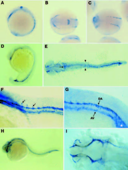
Localization of flk-1 transcripts in wild-type embryos. 7- somite stage (A-C), 20-somite stage (D,E), 24-somite stage (F,G), and 24 hpf (H,I). Embryos shown in A,D,F-H are lateral views with dorsal to the top and anterior to the left. All others are dorsal views with anterior to the left (B shows the head region, C shows the midtrunk region; F and G show the anterior and posterior trunk regions respectively, and I shows the head region). (A-C) flk-1-positive cells are found in discrete bilateral stripes both anteriorly and posteriorly. There is also a transverse ectodermal stripe of staining in the hindbrain region; this staining quickly becomes weaker (see D) and although blood vessels do form there at later stages, the fate of these flk-1-expressing cells is not entirely clear (see also Figs 4 and 5). (D,E) flk-1 expression appears to extend caudally from the head region as well as in both directions in the trunk region, until by the 20-somite stage there is a continuous band of flk-1-expressing cells from the anterior head region to the tailbud. Concurrently, flk-1- expressing cells in the mid- and posterior trunk regions converge medially. The site (arrowheads) where the paired lateral dorsal aortae fuse into a single medial aorta is the site of vascular breakdown in the gridlock mutation (Weinstein et al., 1995). At a slightly later stage, single cells (F, arrows) are starting to migrate into the intersomitic space in the upper trunk region to form the intersomitic vessels. The dorsal aorta (DA) and axial vein (AV) are clearly distinct in the trunk region at this stage, and further caudally, in the tail, there is a region of more intense staining (asterisk). [Similarly intense flk-1 expression is also observed in the tail region of early mouse embryos (Yamaguchi et al., 1993; Shalaby et al., 1995)]. By 24 hpf (H, I), the whole vasculature including the head vessels (I) is clearly outlined by staining for flk-1 transcript.
|
