
Fig. 8
- ID
- ZDB-FIG-140228-38
- Publication
- Schilling et al., 1996 - The chinless mutation and neural crest cell interactions in zebrafish jaw development
- Other Figures
- All Figure Page
- Back to All Figure Page
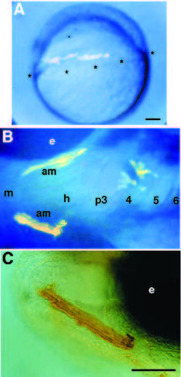
Mutant mesodermal cells form muscles in a wild-type host. All three panels show stages in the development of a single mosaic animal made by transplanting premesodermal cells from a chn donor into a wild-type host at early gastrula stage. (A) 6 hour, lateral view. In the gastrula, rhodamine fluorescence is visible in cells near the equator, at the margin of the blastoderm (asterisks), where they will involute to form mesendoderm. (B) 72 hour, lateral view. Labelled mutant muscle cells are shown in both of the adductor mandibulae muscles in the wild-type mandibular arch of the host. (C) Biotinlabeled, mutant muscle cells in the adductor mandibula. Abbreviations: am, adductor mandibula; e, eye; h, hyoid arch; m, mandibular arch; p3-6, pharyngeal arches 3-6. Scale bars, 100 μm. |