
Fig. 8
- ID
- ZDB-FIG-140227-50
- Publication
- Melby et al., 1996 - Specification of cell fates at the dorsal margin of the zebrafish gastrula
- Other Figures
- All Figure Page
- Back to All Figure Page
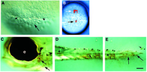
Dorsal marginal blastomeres can contribute to the forerunner cells. (A) Nomarski optics photograph showing the appearance of the forerunner cells at shield stage. Arrow points to the forerunner cell cluster, and arrowheads point to the margin of the blastoderm. B-E are from an embryo in which a labeled marginal blastomere contributed to both anterior mesendoderm and forerunner cell derivatives. (B) Dorsal view of the live embryo at late shield stage. The clone consists of a cluster of cells in the anterior hypoblast (white arrow) and two cells in the forerunner cell population (black arrow). (C-E) Whole-mount side views of the embryo at 3d, after it had been fixed and stained for the lineage label, biotin. Biotin staining appears brown, while pigment cells appear black. (C) The head region, showing labeled pharyngeal endoderm (arrows), derived from cell(s) in the anterior hypoblast. e, eye. (D) Part of the tail showing labeled muscle cells (arrows). (E) More posterior view of the tail showing labeled notochord (arrowhead), body wall mesenchyme (arrows), and fin mesenchyme (small arrow). Scale bar: 40 μm in A; 250 μm in B; 50 μm in C-E. |