
Fig. 2
- ID
- ZDB-FIG-140227-45
- Publication
- Melby et al., 1996 - Specification of cell fates at the dorsal margin of the zebrafish gastrula
- Other Figures
- All Figure Page
- Back to All Figure Page
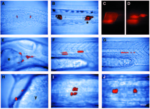
The fates of cells from the dorsal marginal region of the gastrula. (A,E-H) Side views during the early pharyngula period (24-36h); (BD) side views at 3d; (I,J) dorsal views during the pharyngula period. Anterior is to the left in all panels. A-D show the same embryo, in which coin-shaped cells of the notochord rudiment gave rise to both vacuolated notochord cells and notochord sheath by 2d. (A) The clone initially contained three cells at 1d: one cell anteriorly and two posteriorly. (B) At 3d, the anterior cell had become a vacuolated cell, and the posterior cells had generated one vacuolated cell and four notochord sheath cells. (C,D) Higher power fluorescent views of the posterior cells (asterisk in B) at different focal planes at 3d. (C) Vacuolated cell. (D) Sheath cells. Only two of the four notochord sheath cells are in focus. (E) Head mesenchyme: cells that were ventral and lateral to the brain, and were mesenchymal during the early pharyngula period. We sometimes observed these clones differentiating as endothelial cells (n=6) or parachordal cartilage (n=2). Head muscles also arise from cells that are mesenchymal at 1d (Schilling and Kimmel, 1994; Kimmel et al., 1990; Warga, 1996). (F) Somitic muscle: these cells spanned the length of the somite. Muscle clones were confined to one side of the animal if located in the trunk, but were often bilateral if located in the tail (Kimmel and Warga, 1987). (G) Hypochord: an axial tissue lying just below the notochord, and extending from the level of the first somite through the tail. Like the floor plate of the spinal cord, hypochord is composed of a single midline row of cells in zebrafish (Hatta and Kimmel, 1993). (H) Hatching gland: large granule-filled cells that lie on the surface of the pericardium. (I) Ventral nervous system: these clones included both morphologically identifiable neurons and other cells presumed to be glia or neuroblasts. This clone excluded floor plate. Neuroepithelial cells such as these and floor plate cells have a similar cuboidal morphology in side views, but floor plate can be distinguished in a dorsal view as forming a single midline row of cells. (J) Floor plate: part of a clone derived from a deep cell in the third layer of the epiblast. A,B,E-J are composites of bright-field and fluorescent images taken at the same focal plane; C and D are fluorescent images only. Scale bar, 40 μm in A,B,E-H; 100 μm in C,D,I,J. |