
Fig. 8
- ID
- ZDB-FIG-130905-18
- Publication
- Filipek-Górniok et al., 2013 - Expression of chondroitin/dermatan sulfate glycosyltransferases during early zebrafish development
- Other Figures
- All Figure Page
- Back to All Figure Page
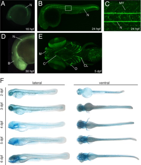
The notochord and cartilage structures are sites of CS/DS accumulation. A–D: Lateral views of zebrafish embryos and larvae showing CS deposition as detected with the CS-56 antibody. The notochord is weakly stained at 16 hpf (A) and more prominent CS deposition is seen at 24 hpf and 30 hpf (B–D). CS deposition in the myoseptum is shown in C, which is a magnification of the boxed area in B. Brain tissues are weakly stained at 30 hpf (D). E: CS is present in the pharyngeal cartilages (the Meckel′s cartilage and the ceratohyal) and in dermal bones (the operculum and the cleithrum) at 5 dpf. F: Ventral and lateral views of a zebrafish embryo stained with Alcian blue to detect CS/DS at 2–6 dpf. B, brain; C, ceratohyal; CL, cleithrum; M Meckel′s cartilage; MY, myoseptum; N, notochord; O, operculum. |
| Antibody: | |
|---|---|
| Fish: | |
| Anatomical Terms: | |
| Stage Range: | 14-19 somites to Day 5 |