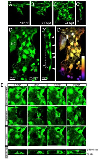
ECs of the CCV delaminate from the migrating sheet and align around the lumen. Confocal projections from time-lapse movies of a Tg(etsrp:EGFP)ci1 (A-C′,E) or a Tg(fli1a:EGFP)y1 (D-D′) embryo, dorsolateral views. (A-D′) At 20 hpf, ECs start to migrate to form a monolayer on top of the YSL (A-C′). Individual ECs delaminate from the YSL side and thereby align around a lumen (D′,E). At 26 hpf, an initial lumenized open-ended cylinder is formed (D-D′). This cylinder becomes extended by the ECs underneath the epidermis (arrowheads in D′). Sagittal sections based on 3D reconstructions of C and D (at the levels indicated by the dashed lines) show a monolayer and no lumen (C′) and a lumen (D′). Arrowheads in D′ point to the ECs migrating to extend the CCV. (D′) Depth color coding of the confocal maximum projection shown in D. Proximal-most structures are white, whereas darker colors represent more distal structures. (E) Delamination of a single EC (asterisk) from the endothelial monolayer on top of the YSL to the epidermal side of the CCV. To visualize this process, the second and third panels contain only part of the maximum projection: in the panels labeled ‘epidermal side’, ECs located on the YSL side are cropped away, whereas in the panels labeled ‘YSL side’, ECs located on the epidermal side are cropped away. NC, neural crest; BC, blood cells; YSL, yolk syncytial layer.
|
