FIGURE

Fig. S2

ID
ZDB-FIG-130618-14
Publication
Villefranc et al., 2013 - A truncation allele in vascular endothelial growth factor c reveals distinct modes of signaling during lymphatic and vascular development
Other Figures
All Figure Page
Back to All Figure Page
Fig. S2

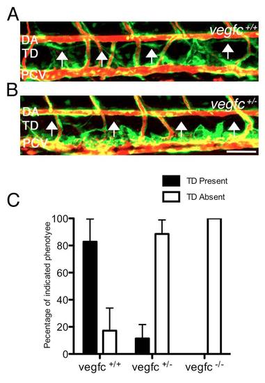
Decreased thoracic duct formation in vegfcum18 heterozygous embryos. (A,B) Confocal microangiographs of trunk blood vessels in Tg(fli1a:egfp)y1 larvae at 5 dpf in progeny from a um18 carrier incross. Microangiography dye is pseudocolored red. (A) Normal TD formation in a wild-type embryo. (B) Loss of TD in vegfcum18/+ heterozygous embryo. The normal location of the thoracic duct (TD) is indicated by arrows. Lateral view, dorsal is up and anterior to the left. DA, dorsal aorta; PCV, posterior cardinal vein. (C) Quantification for presence of TD in embryos of the indicated genotype. Scale bar: 25 μm.

Expression Data

Expression Detail
Antibody Labeling
Phenotype Data
Fish:
Observed In:
Stage: Day 5

Phenotype Detail
Acknowledgments
This image is the copyrighted work of the attributed author or publisher, and ZFIN has permission only to display this image to its users. Additional permissions should be obtained from the applicable author or publisher of the image. Full text @ Development