
Fig. 2
- ID
- ZDB-FIG-130516-39
- Publication
- Lévesque et al., 2013 - Inflammation drives wound hyperpigmentation by recruiting pigment cells to sites of tissue damage
- Other Figures
- All Figure Page
- Back to All Figure Page
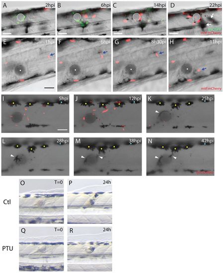
Both melanoblasts and melanocytes are recruited to the wound. (A-D) Stills taken from supplementary material Movie 2 showing neutrophils and melanoblasts as they migrate to a bead implanted in a lysC:GFP/mitf:gal4-UAS:mCherryfish larva. (A) Neutrophils (lysCGFP, green) are rapidly recruited to the wound from 2 hours after implantation. (B) By 6 hours after wounding, melanoblasts (mitf:Gal4-UAS:mCherry, red) arrive close to the bead. (C) This continues even as the inflammatory response is resolving at 14 hpi. (D) By 22 hours, melanocytes have also migrated to the wound (out of focus, indicated by white arrows). (E-H) Stills taken from supplementary material Movie 3 showing activation and migration of an individual melanoblast (mitf:mCherry, red and highlighted by a blue arrow) towards the bead. (I-N) Stills taken from supplementary material Movie 4 showing the migration of both neutrophils (red) and melanocytes (black) to the wound (bead has been implanted in a lysC:DsRed fish larva). (I) Number of neutrophils (lysC:DsRed, red) peak at the wound several hours prior to any observable response by melanocytes (yellow and blue dots). (J) First indications of melanocyte response to the wound commence at about 12 hpi (only those with yellow dots), before the final resolution of neutrophils. (K) Several melanocytes make slow progress towards the bead and by 25 hpi a smaller melanocyte has also arrived and is in close contact with the bead (white arrowhead). (L-N) Movement of small and more mature dorsal melanocytes continues for many hours, with cells extending long protrusions to reach the bead. (OR) Comparison of fish treated with PTU at the time of bead implantation versus untreated controls. In both control (O,P) and PTU-treated fish (Q,R) the wound is pigmented by 24 hpi. Scale bars: 50 μm (A-R). |