
FIGURE
Fig. 3
- ID
- ZDB-FIG-130514-6
- Publication
- Wages et al., 2013 - Changes in zebrafish (Danio rerio) lens crystallin content during development
- Other Figures
- All Figure Page
- Back to All Figure Page
Fig. 3
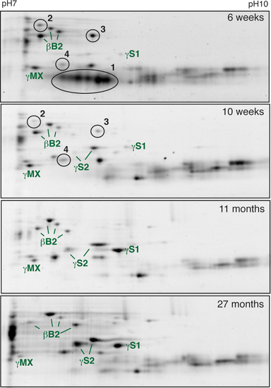
Separation of total zebrafish lens protein using 7–10 pH strips identifies ontogenetic changes in β/γ-crystallin expression. The identities of several crystallins found in each gel are noted. Numbered ovals indicate protein spots found only in the 6- and 10-week lenses. |
Expression Data
Expression Detail
Antibody Labeling
Phenotype Data
Phenotype Detail
Acknowledgments
This image is the copyrighted work of the attributed author or publisher, and
ZFIN has permission only to display this image to its users.
Additional permissions should be obtained from the applicable author or publisher of the image.