Fig. 1
- ID
- ZDB-FIG-130411-28
- Publication
- Veerkamp et al., 2013 - Unilateral dampening of bmp activity by nodal generates cardiac left-right asymmetry
- Other Figures
- All Figure Page
- Back to All Figure Page
|
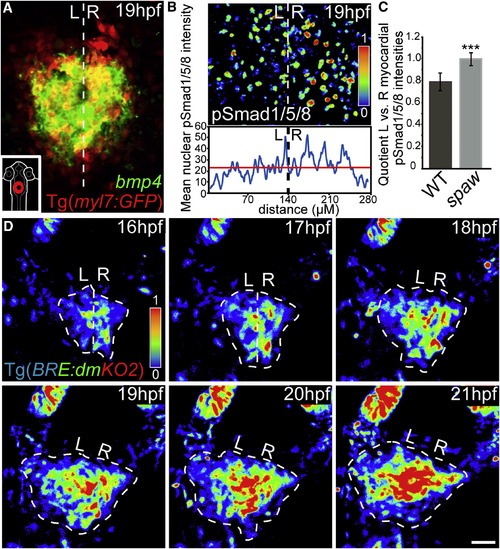
Bmp Signaling Is Weaker within the Left Heart Cone prior to the Appearance of Morphological Asymmetries(A) Fluorescence two-color in situ hybridization of bmp4 expression at the heart cone stage (false-colored image).(B) Cardiac progenitor cells have lower nuclear pSmad-1/5/8 intensities on the left side of the heart as indicated by the color range. The mean intensity plot is a measure of the entire intensities within the field of view.(C) The L/R asymmetry of nuclear pSmad-1/5/8 intensities is abolished in spaw morphant embryos at 19 hpf (mean with SD based on quantifications of eight hearts per genotype; p < 0.0005).(D) The laterality of cardiac jogging coincides with lower intensities of Bmp signaling activity as indicated by the Bmp reporter line Tg[BRE-AAVmlp:dmKO2]mw40 (false-colored image). White dotted line indicates the embryonic midline. Scale bar, 50 μm. L, left; R, right.See also Figures S1, S2, and Movie S1. |
| Genes: | |
|---|---|
| Antibody: | |
| Fish: | |
| Knockdown Reagent: | |
| Anatomical Terms: | |
| Stage Range: | 14-19 somites to 20-25 somites |
| Fish: | |
|---|---|
| Knockdown Reagent: | |
| Observed In: | |
| Stage: | 20-25 somites |
Reprinted from Developmental Cell, 24(6), Veerkamp, J., Rudolph, F., Cseresnyes, Z., Priller, F., Otten, C., Renz, M., Schaefer, L., and Abdelilah-Seyfried, S., Unilateral dampening of bmp activity by nodal generates cardiac left-right asymmetry, 660-667, Copyright (2013) with permission from Elsevier. Full text @ Dev. Cell

