
Fig. 2
- ID
- ZDB-FIG-130326-36
- Publication
- Uribe et al., 2013 - Dimethyl Sulfoxide (DMSO) Exacerbates Cisplatin-induced Sensory Hair Cell Death in Zebrafish (Danio rerio)
- Other Figures
- All Figure Page
- Back to All Figure Page
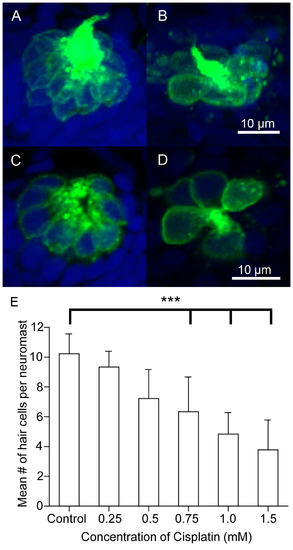
Dose response curve following cisplatin treatment. Five-days post-fertilization Brn3c-GFP transgenic zebrafish were exposed to varying doses of cisplatin for four hours to determine at which dose approximately 50% of the hair cells die. The larvae were fixed, co-labeled with the nuclear dye TO-PRO-3 (blue), and the GFP-tagged hair cells (green) in the O2 neuromast were imaged using confocal microscopy. (A–B) Z-stack projections of two O2 neuromasts under different treatment conditions showing the entire neuromast structure. (C–D) Slices from the same neuromasts as in A, B demonstrating the membrane-bound GFP label surrounding the nuclear dye. (A, C) Hair cells appear normal in untreated controls. (B, D) Noticeably fewer hair cells are found in larvae treated with 1 mM cisplatin. (E) The mean number of hair cells per O2 neuromast (± SD) decreased as the dose of cisplatin increased when compared to untreated controls. n = 931 neuromasts for each treatment group. ***p<0.001 when individual treatments are compared to untreated controls. |