
- Title
-
Dimethyl Sulfoxide (DMSO) Exacerbates Cisplatin-induced Sensory Hair Cell Death in Zebrafish (Danio rerio)
- Authors
- Uribe, P.M., Mueller, M.A., Gleichman, J.S., Kramer, M.D., Wang, Q., Sibrian-Vazquez, M., Strongin, R.M., Steyger, P.S., Cotanche, D.A., and Matsui, J.I.
- Source
- Full text @ PLoS One
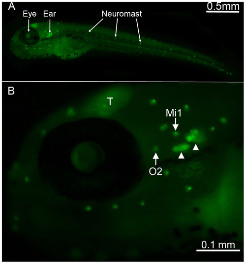
Location of neuromasts on Brn3c-GFP transgenic zebrafish. (A) Lateral view of a 5 days post-fertilization transgenic zebrafish showing GFP expression in neuromasts that are found along the head and the body (bright dots) of the animal. (B) Higher magnification of the head region, with neuromasts containing brightly GFP-labeled hair cells (white arrows) and inner ear organs (white arrowheads) easily identifiable. The otic 2 (O2) and middle 1 (Mi1) neuromasts are highlighted as these are the two neuromasts from which data for this study were obtained. The zebrafish optic tectum (T) is another structure that is also labeled with GFP. |
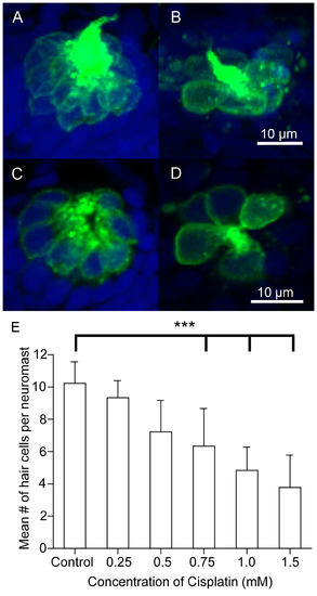
Dose response curve following cisplatin treatment. Five-days post-fertilization Brn3c-GFP transgenic zebrafish were exposed to varying doses of cisplatin for four hours to determine at which dose approximately 50% of the hair cells die. The larvae were fixed, co-labeled with the nuclear dye TO-PRO-3 (blue), and the GFP-tagged hair cells (green) in the O2 neuromast were imaged using confocal microscopy. (A–B) Z-stack projections of two O2 neuromasts under different treatment conditions showing the entire neuromast structure. (C–D) Slices from the same neuromasts as in A, B demonstrating the membrane-bound GFP label surrounding the nuclear dye. (A, C) Hair cells appear normal in untreated controls. (B, D) Noticeably fewer hair cells are found in larvae treated with 1 mM cisplatin. (E) The mean number of hair cells per O2 neuromast (± SD) decreased as the dose of cisplatin increased when compared to untreated controls. n = 931 neuromasts for each treatment group. ***p<0.001 when individual treatments are compared to untreated controls. |

Co-treatment with DMSO and cisplatin compromises hair cell morphology and kills hair cells. Zebrafish were treated with embryo medium (control), 0.5% DMSO, two different concentrations of cisplatin (0.5 mM or 1 mM), or cisplatin (0.5 mM or 1 mM) with 0.5% DMSO. Representative images of (A) control neuromasts, (B) neuromasts exposed to 1 mM cisplatin, and (C) neuromasts exposed to 1 mM cisplatin and 0.5% DMSO. When compared to a control O2 neuromast (A), fewer hair cells were present in the O2 neuromast of a 1 mM cisplatin-treated larva (B). When cisplatin is co-incubated with 0.5% DMSO (C), even fewer hair cells remain within the neuromast. (D) When larvae were treated with 0.5% DMSO in conjunction with cisplatin, there was a significant reduction in the number of hair cells when compared to cisplatin treatment alone indicating a synergistic effect of DMSO and cisplatin. Results are the mean values ± SD. n = 1351 neuromasts for each treatment group. ***p<0.001. |

More Texas Red fluorescence in hair cells when DDP-TR is solubilized in DMSO than in methanol. Brn3c-GFP zebrafish were treated with 2 µg/mL DDP-TR dissolved in either methanol or 0.1% DMSO for two to forty-eight minutes and then fixed and imaged. (A) Low levels of DDP-TR fluorescence (red) were found in the O2 hair cells of methanol-treated zebrafish after two minutes. (B) More DDP-TR fluorescence was present in the hair cells of 0.1% DMSO-treated animals after two minutes. (C) DMSO increased levels of DDP-TR fluorescence into neuromast hair cells over time compared to DDP-TR plus methanol-treated fish. Results are the mean values ± SD. n = 15125 individual hair cells per time point per treatment. **p<0.01. |