|
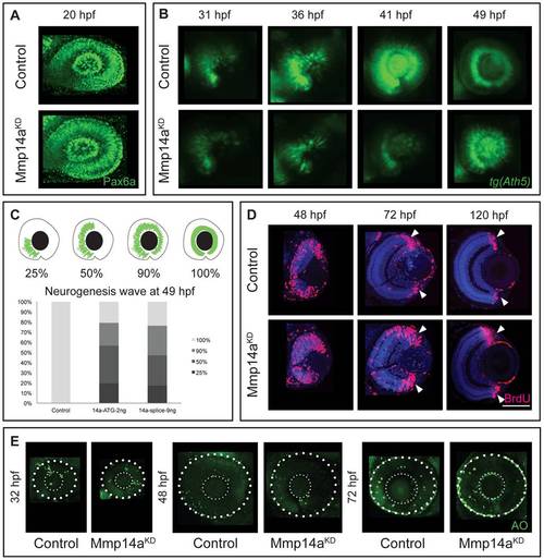
Mmp14a knockdown affects neurogenesis in the retina. A Whole mount immunostaining for Pax6a does not reveal any differences in eye morphology and Pax6a expression between Mmp14a morphant and control retinas at 20 hpf. B Live imaging of retinal neurogenesis in transgenic Tg(Ath5:GFP) embryos shows a small patch of Ath5+ cells in the ventronasal region of the developing eye in control embryos at 31 hpf. The Ath5+ cell population expands in a wave and reaches the ventrotemporal region of the retina around 41 hpf. Mmp14a morphant eyes show a delay in neurogenesis and the Ath5+ cell population only reaches the ventrotemporal region of the retina around 49 hpf. C The progression of the expansion of the Ath5+ cell population at 49 hpf is semi-quantitatively scored as illustrated in the top scheme. Injection of both the Mmp14a-ATG and the Mmp14a-splice MOs resulted in a severe delay of the neurogenesis wave (n = 45 from 3 independent experiments). D BrdU incorporation assays, performed at various developmental stages, show a higher number of immunopositive BrdU (BrdU+) cells in transverse sections of morphant retinas at 48 hpf. At 72 hpf, the proliferating region is restricted to the CMZ (white arrowhead) in control retinas, whereas BrdU+ cells are still abundantly detected in the central retina of Mmp14a morphant eyes. Even at 120 hpf, the proliferating region in morphant retinas is less confined to the CMZ as compared to control retinas. DAPI (blue) was used as counterstain. E Labeling of apoptosis using the fluorescent marker acridine orange does not reveal significant differences in the number of apoptotic cells at various developmental stages between Mmp14a morphant and control retinas. The small dotted circle marks the lens, the larger circle lines the eye (n = 20 from 4 independent experiments). AO, acridine orange; CMZ, cilliary marginal zone; hpf, hours post fertilization. Scale bar in D: 50 μm.
|