|
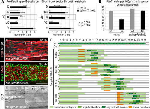
Induced Tbx6 expression differentially rescues central dermomyotome and segmentation. (A) Dorsal/ventral distribution of proliferative (phosphorylated Histone H3, pH3+) cells in non-tg and tg(hsp70:tbx6) wild type sibling and fss/tbx6 mutant embryos 8hours after heat-shocking at the 8S stage. Data are presented as mean ± s.e.m. (B) Graph showing number of Pax7+ cells in non-tg and tg(hsp70:tbx6) wild type sibling and fss/tbx6 mutant embryos at 10hours after heat-shocking at the 10S stage. (C–E) Rescue of dermomyotome and segmentation in fss/tbx6 mutant tg(hsp70:tbx6) visualized using F59 (red) and MF20 (white) (C) and Pax7 (green) and Mef2 (white) (D) labeling, and DIC imaging (E). Specimen stages are 5d (C,E) and 24h (D), rescued segments are numbered according to the corresponding region in wild-type siblings. (F) Schematic showing the location of rescued dermomyotome (light green), segments (dark green) in individual embryos after heat shocks at various times (orange). Note that a Tbx6 pulse rescues the formation of 1–3 somite boundaries and central dermomyotome over 8–10 somite lengths. Scale bars: 100μm (C,D), 50μm (E).
|