|
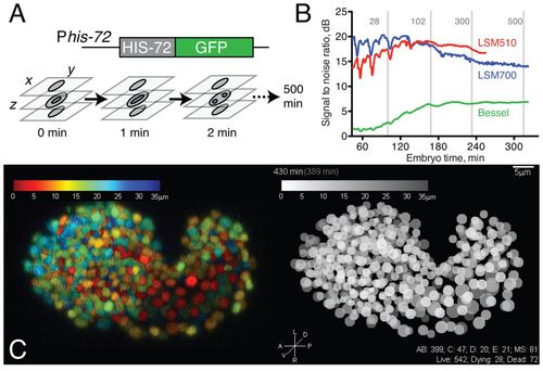
4D data acquisition and image properties. (A) Schematic of acquisition setup. We use laser scanning confocal microscopy to acquire z-stacks every minute for 5 hours of embryo development followed by z-stacks every two minutes for 3 hours at 20-24°C. Acquisition of each stack takes 12-15 seconds at 512 × 275 × 35 voxels. (B) Comparison of signal-to-noise ratio (SNR) of raw data from 4D movies taken using Zeiss LSM510, Zeiss LSM700 and Bessel beam microscopy. Image acquisition was optimized for image quality compatible with viability; under these parameters, the LSM510 and LSM700 data sets showed comparable SNR. (C) Comparison of confocal image (color depth code, projection of LSM700 z-stack, 430 minutes) with digital representation (gray scale depth code).
|