
FIGURE
Fig. 2
Fig. 2
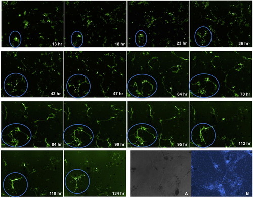
Differentiation of flk1-GFP-Labeled Endothelial Cells in Primary Culture Flk1-GFP expressing endothelial cells are scattered at day 1 and become flatten and elongated at days 2–4, undergoing tube formation. After 5–6 days of culture, GFP-positive cells start to die (blue circles). (A and B) Primary cells stained by (A) DIC and (B) Hoechst 33342 (5 μg/ml for 4.5–5 hr) on day 6. See also Figures S1 and S2. |
Expression Data
Expression Detail
Antibody Labeling
Phenotype Data
Phenotype Detail
Acknowledgments
This image is the copyrighted work of the attributed author or publisher, and
ZFIN has permission only to display this image to its users.
Additional permissions should be obtained from the applicable author or publisher of the image.
Full text @ Cell Rep.