
Fig. 5
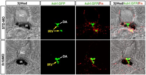
The effect of fn1b knockdown on the interrenal tissue and the peri-interrenal vasculature. Transverse sections of Tg(kdrl:EGFP)s843 embryos were injected with either STD-MO or fn1b antisense morpholino, harvested at 3 dpf and visualized for 3β-Hsd activity (black), GFP (green) and Fn expression (red). All sections are oriented with the posterior end toward top of page. The IRV structure (yellow arrows) of a 3 dpf control embryo appeared discontinuous on a single confocal section because the IRV at this stage, after spanning through the interrenal tissue (IR), curved anteriorward and then posteriorward before connecting to the AMA segment. Fn doposition in the interrenal region, as well as the interrenal migration and angiogenesis, remain unperturbed in the fn1b morphant. The Fn expression and IRV morphology in the fn1b morphant shown here is a representative of 10 samples exhibiting a disruption of somite boundaries characteristic of the Fn1b-deficient embryo. Abbreviations: notochord (NC), interrenal tissue (IR), dorsal aorta (DA), interrenal vessel (IRV). |
| Genes: | |
|---|---|
| Fish: | |
| Knockdown Reagents: | |
| Anatomical Terms: | |
| Stage: | Protruding-mouth |
| Fish: | |
|---|---|
| Knockdown Reagents: | |
| Observed In: | |
| Stage: | Protruding-mouth |