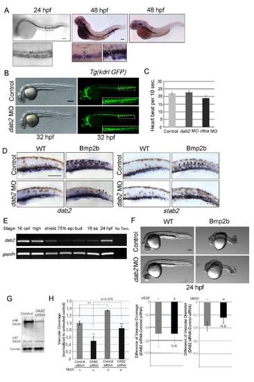
Expression pattern of dab2 in zebrafish embryo and its function in vascular development (related to Figures 1).
(A) Micrographs of 24hpf wild-type zebrafish embryos showing dab2 expression. Initially, dab2 is expressed in both arterial and venous endothelial cells. Area within rectangle is shown in higher magnification. Scale bar is 100μm. At 48hpf, dab2 is strongly expressed within the otic vesicle, pronephric duct, and venous endothelial cells. Areas within rectangles are shown in higher magnification. Scale bar is 200μm. Homozygous cloche embryos, which lack a majority of endothelial cells, only retain dab2 expression within the otic vesicle and pronephric duct (right). Abbreviations: A: aorta, O: otic vesicle, P: pronephric duct, V: vein. (B) Micrographs of control or dab2 MO injected embryos. Inhibition of Dab2 does not cause any discernible morphological defects in early stages. The most pronounced phenotypic defects were localized within the developing CVP. Areas within white rectangles are shown in a higher magnification as insets. Scale bar is 200μm. (C) Heart beats in dab2 and cltca MO injected embryos. Dab2 and Cltca deficient embryos have normal heart beat at 48hpf. (D) Micrographs of 45hpf wild-type or Tg(hsp70l:bmp2b) embryos injected with control or dab2 MO. Ectopic vessels in Bmp2b over-expressing embryos were strongly stained with venous specific marker dab2 and stab2, indicating the venous nature of these vessels. Arrows point the ectopic vessels. Scale bar is 200μm. (E) Expression of dab2 message during development measured by semi quantitative RT-PCR, demonstrating that dab2 is maternally provided. (F) Micrographs of 24hpf wild-type or Tg(hsp70l:bmp2b) embryos injected with control or dab2 MO which were heat-shocked at 10 hpf to induce over-expression of Bmp2b. Blocking Dab2 did not alleviate Bmp2b induced anterior-posterior axis defects, suggesting that Dab2 may mediate Bmp2b signaling in a stage specific manner. Scale bar is 100μm. (G) Validation of DAB2 siRNA in HUVEC. (H) Effects of DAB2 inhibition on VEGF-A mediated angiogenic responses in HUVECs. Control or DAB2 siRNA treated cells were incubated in the presence or absence of VEGF-A on Fibrin gel. Total vascular coverage and relative changes in the vascular coverage and diameter upon VEGF-A treatment in control or DAB2 siRNA-treated HUVECs were quantified by PRISM software. N=3; N.S, no significance; *p<0.05 and **p<0.005. Error bar presents standard error in the mean.
|
