
Fig. 2
- ID
- ZDB-FIG-120717-19
- Publication
- Gaudin et al., 2012 - Chemoattractant axon guidance cues regulate de novo axon trajectories in the embryonic forebrain of zebrafish
- Other Figures
- All Figure Page
- Back to All Figure Page
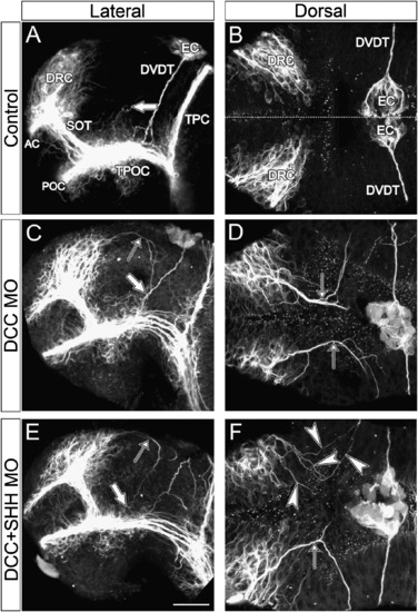
dcc loss-of-function generates an ectopic axon bundle in the diencephalon. All images are confocal projections of zebrafish whole-mounted brains at 30 hpf immunolabelled for expression of acetylated α-tubulin. Anterior is left in all images. (A) The axon tracts are displayed according to Fig. 1. Note the absence of axons in the diencephalic territory between the DVDT and DRC–SOT except for a few pioneers of the future THC (large arrow). (B) Note the absence of any axon tract in the diencephalic space separating the DRC from the EC. The dotted line represents the midline. (C) Axons emerging from the posterior edge of the DRC and extending into the diencephalon are indicated by the thin arrows. These axons typically follow a curved route through the diencephalon down to the TPOC, meeting with the pioneers of the THC extending dorsally or the DVDT (large arrow). (D) The ectopic axons clearly extend from the caudal margins of the DRC into the dorsal diencephalon. Most axons are tightly bundled at the exit of the telencephalic cluster. (E) Co-knockdown of shh and dcc did not rescue the phenotype of these ectopic diencephalic axons nor did it alter their route ventrally (arrow) since they still connect with the TPOC (large arrow). (F) While the left side shows a typical tightly-fasciculated bundle of ectopic axons (arrow), the right side of this embryo exhibits axons wandering individually in the dorsal diencephalon (arrowheads). These individual axons appear to be still following a stereotypical route as in the contralateral side of the brain. Scale bar=35 μm (A, C, E), 20 μm (B, D, F). |
Reprinted from Developmental Biology, 367(2), Gaudin, A., Hofmeister, W., and Key, B., Chemoattractant axon guidance cues regulate de novo axon trajectories in the embryonic forebrain of zebrafish, 126-139, Copyright (2012) with permission from Elsevier. Full text @ Dev. Biol.