
Fig. S3
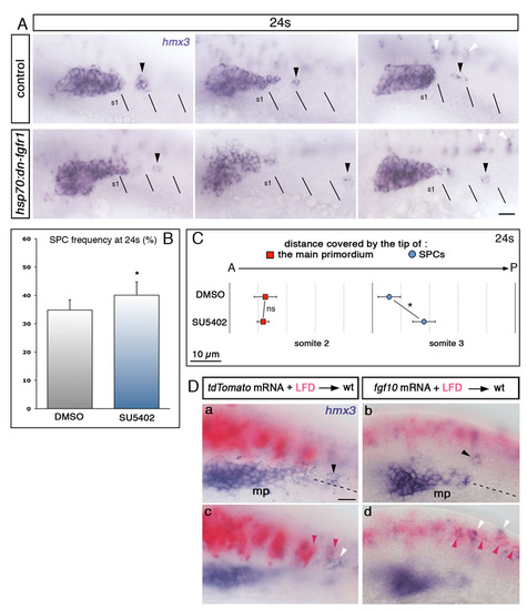
Fgf signalling attracts SPCs towards the primordium. Side view and anterior to the left. (A) hmx3 expression in heat-shocked hsp70:dn-fgfr1 embryos and control siblings. The position of SPCs, indicated by black arrowheads, is shifted caudally in hsp70:dn-fgfr1 embryos. Black lines indicate somitic borders. White arrowheads indicate hmx3-expressing spinal cord neurons. (B) SPC frequency at 24s in DMSO- and SU5402-treated embryos. *P=0.018. (C) Average distances covered by the tip of the main primordium (red) and SPCs (blue) in SU5402-treated embryos (n=49) and DMSO controls (n=34) at 24s. *P=0.041, ns, non significant (P=0.85). (D) Additional examples of hmx3 expression (purple) in embryos transplanted with Fgf10- or tdTomato-expressing cells (pink). Black, pink and white arrowheads show SPCs, transplanted cells, and hmx3-expressing spinal cord neurons, respectively. Dashed lines represent the horizontal somitic midlines. Focal planes focused on the primordium and SPCs (a,b) or on the transplanted cells in the neural tube (c,d). mp, main primodium; S, somite. Scale bars: 25 μm. |