
Fig. S3
- ID
- ZDB-FIG-120601-22
- Publication
- Chablais et al., 2012 - The regenerative capacity of the zebrafish heart is dependent on TGFβ signaling
- Other Figures
- All Figure Page
- Back to All Figure Page
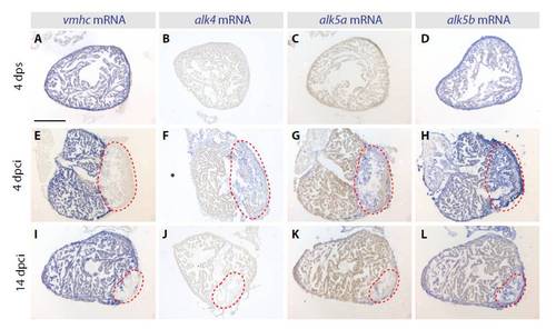
Expression patterns of TGFβ/Activin type I receptors in the cryoinjured hearts. In situ hybridization of consecutive heart sections with vmhc, alk4, alk5a and alk5b mRNA antisense probes (blue staining) at different phases of heart regeneration. (A-D) In uninjured sham operated hearts at 4 dps, vmhc (A) is expressed in the entire ventricle, whereas alk4 (B) and alk5a (C) are not detected. alk5b (D) is ubiquitously expressed in the ventricle. (E-L) In regenerating hearts at 4 and 14 dpci, the absence of vmhc demarcates the injury site (red dashed lines). alk4 and alk5a are weakly induced in the post-infarct area. alk5b is expressed in both the intact myocardium and in the post-infarct. Scale bar: 300 μm. |