
Fig. 3
- ID
- ZDB-FIG-120130-3
- Publication
- Schuhmacher et al., 2011 - Evolutionary relationships and diversification of barhl genes within retinal cell lineages
- Other Figures
- All Figure Page
- Back to All Figure Page
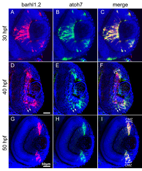
Double fluorescent in situ hybridization of barhl1.2 and atoh7. Confocal sections through the central retina of embryos hybridized with barhl1.2 (A, D and G, in red) and atoh7 (B, E and H, in green) antisense RNA probes. (C, F and I) merge of red and green channels. Nuclei were stained with DAPI (blue). View is frontal in all pictures, anterior is always to the top. Stages analyzed are indicated. (D- 25 -F) Downregulation of barhl1.2 in the central retina is delayed with respect to the one of atoh7 but overlapping in the ciliary marginal zone (highlighted with white brackets CMZ). White arrows highlight two cells where both barhl1.2 and atoh7 are expressed. (F and I) the white brackets indicate the CMZ where barhl1.2 and atoh7 expression always overlap. |
| Genes: | |
|---|---|
| Fish: | |
| Anatomical Terms: | |
| Stage Range: | Prim-15 to Long-pec |