
Fig. 7
- ID
- ZDB-FIG-111220-34
- Publication
- Chatterjee et al., 2011 - Conserved And Non-Conserved Enhancers Direct Tissue Specific Transcription In Ancient Germ Layer Specific Developmental Control Genes
- Other Figures
- All Figure Page
- Back to All Figure Page
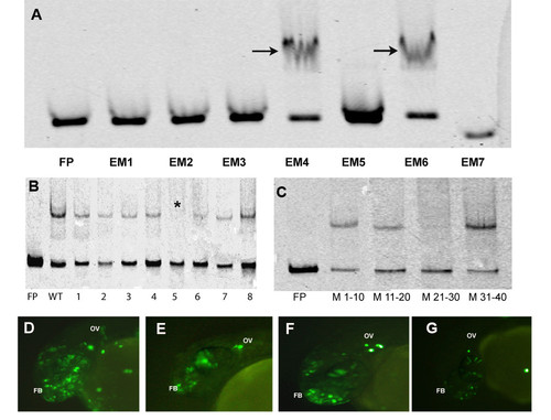
EMSAs and transgenic assays to detect core-binding motif. (A) Electrophoretic Mobility Shift Assays (EMSA) with 7 overlapping probes spanning the 200 bp non-conserved enhancer. Arrows indicate the two probes that bound protein. (B) EMSAs with 5 bp sliding mutant probes for EM4. The star indicates the probe 5 which did not show binding and the subsequent probe 6 showed weak binding. (C) EMSA with 10 bp mutation probes. Mutations in nucleotides 21-30 (M21-30) leads to complete abrogation of binding. Transgenic assays with wild type 40 bp probe (EM4) (D), mutant probe 5 (E), mutant probe 6 (F) and 10 bp mutant probe (G). The enhancer could drive expression in forebrain (FB) and otic vesicle (OV) at 24 hpf that are domains of expression of otx1b. |